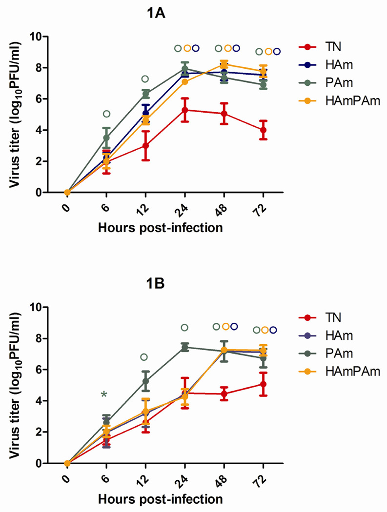
Figure 1.
Replication of rg-A/Tennessee/560/09 (TN/09, in red), rg-A/Tennessee/560/09-HA K154Q mutant (HAmut, in blue), rg-A/Tennessee/560/09-PA L295P mutant (PAmut, in green), and rg-A/Tennessee/560/09-HA K154Q mutant and PA L295P mutant (HAmutPAmut, in orange) in NHBE cells. NHBE cultures were infected via the apical side with each virus at an MOI 0.1 at either 37°C (A) or 33°C (B). The progeny viruses released from the apical surfaces of infected cultures were collected at the indicated time points and titrated in MDCK cells by performing a plaque assay. Representative results expressed as log10 numbers of PFU/ml from two independent experiments are shown. * P < 0.05, ° P < 0.01 compared with the value for TN/09 virus (one-way ANOVA).

