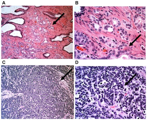
Fig. 1.
Distinct histologic features of prostatic adenocarcinoma and SCNC. The upper panel (A,B) are low and high power pictures of prostatic adenocarcinoma with cancerous glands recapitulating the morphologic features of normal prostatic glands/ducts, and the tumor cells forming glandular structures. Note that many tumor cells show prominent nucleoli. The lower panel (C,D) are low and high power pictures of prostatic SCNC with high-grade neuroendocrine morphology including diffuse, solid growth pattern, high N/C ratio, fine nuclear chromatin pattern, and frequent mitotic figures. Note that there is no glandular formation and no prominent nucleoli (H&E100× (A,C) and 400× (B,D)). [Color figure can be viewed in the online issue, which is available at wileyonlinelibrary.com.]

