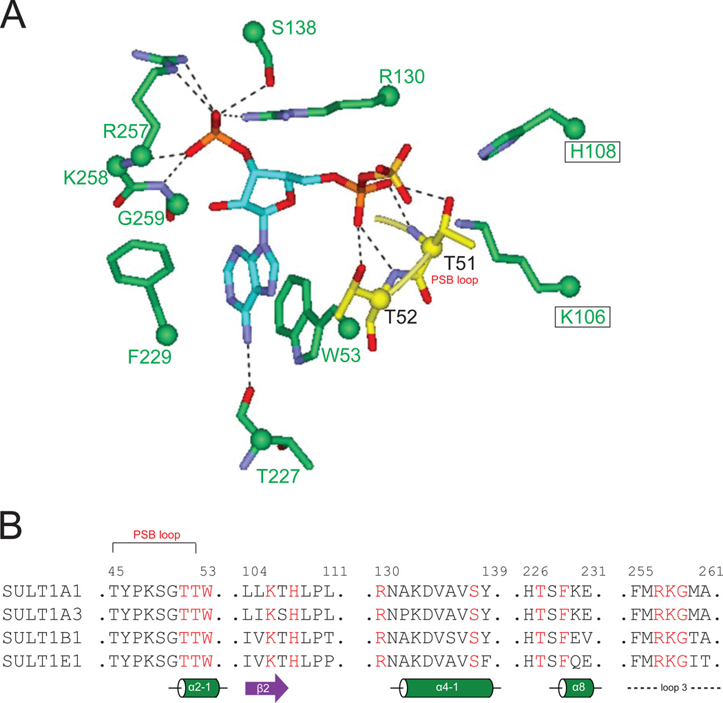
Figure.4. Conserved interaction of PAPS with SULT residues.
Panel A: The conserved PAPS interacting residues from the four SULT crystals, showing their interactions with the PAPS. Dashed lines indicate hydrogen bonds. Panel B: Alignment of four SULT1 sequences showing the PAPS-interacting residues (in red) in secondary structures. Residue numbers in SULT1A1 are labeled,

