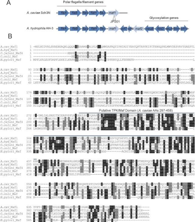
Figure 1.
(A) Genetic organization of the polar flagella loci of Aeromonas caviae Sch3N and A. hydrophila AH-3. Genes known to be involved in flagella filament formation or glycosylation are blue and maf genes are gray. The location of kanamycin resistance marker for the generation of the JPS01 insertion mutant (A. caviae maf1-) is indicated. (B) Alignment of A. caviae Maf1 with homologous Mafs from A. hydrophila AH-3 (accession ABA01574), Campylobacter jejuni subsp. doyley 269.97 (accession YP_001397580), C. coli JV20 (ZP_07400781), and Helicobacter pylori 83 (accession AEE70107). The conserved TPK/Maf domain corresponding to A. caviae Maf1 amino acids 297–458 is highlighted.

