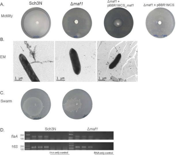
Figure 2.

(A) Analysis of motility of the Aeromonas caviae maf1 mutant JPS01 and derivative strains. Motility as assessed by swimming in 0.25% semisolid motility agar for A. caviae Sch3N (WT), JPS01 (maf1 mutant), and JPS01 containing pBBR1MCS_maf1 and pBBR1MCS. (B) Transmission electron microscopy of the A. caviae strains Sch3N (wild type), maf1-, and maf1- + pBBR1MCS_maf1 grown at 37°C in brain heart infusion broth (BHIB). Bacteria were spotted onto Formvar-coated copper grids and negatively stained using 1% phosphotungstate. (C) Analysis of swarming motility of the maf1 mutant as assessed by movement across the surface of swarming agar. (D) Reverse transcriptase PCR (RT-PCR) analysis of flaA gene expression of A. caviae Sch3N (WT) and maf1-. Primers internal to 16S rRNA gene of A. caviae were used as a control. Experiments were performed in triplicate. Primer pairs are listed in Table 2.
