Fig. 7.
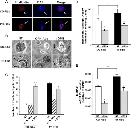
OPN expression levels modulate proinvasive capabilities of PH- and CO-Fibs in 3D Matrigel assays. A: cells were plated onto growth factor-reduced Matrigel in serum-free medium. After 9 days, immunostaining for actin (via Alexa-594-conjugated phalloidin, red) to define cell morphology, and for cell nuclei (DAPI, blue) was performed. B: in 3D growth factor-reduced Matrigel cultures PH-Fibs generated visible filopodia-like structures, a hallmark of cell invasion into Matrigel (arrow in NT image), as opposed to CO-Fibs. In the presence of OPN-specific antibodies (7.5 μg/ml), generation of filopodia in PH-Fibs was inhibited. Addition of rOPN (20 ng/ml) resulted in generation of filopodia (arrows) not only in PH-Fibs, but even in CO-Fibs that did not occur under NT conditions. C: quantification of filopodia-like structures (the number of branch points) was performed via ibidi software analysis: http://www.ibidi.com (Munich, Germany). **P < 0.01, *P < 0.05. D: in Matrigel-coated Boyden chamber assay of cell invasion (see methods), siRNA3-mediated knockdown of OPN expression resulted in reduced invasion through Matrigel (used in a growth factor-reduced form) in both CO- and PH-Fibs. Data represent mean ± SE from 3 different experiments. *P < 0.05 vs. NT cells. E: under NT conditions, MMP-2 expression in PH-Fibs was significantly higher than in CO-Fibs. Treatment of both CO- and PH-Fibs with OPN-siRNA (50 nM, 48 h) resulted in significant decrease of MMP-2 expression (6-fold in CO-Fibs and 4-fold in PH-Fibs) compared with respective NT cells.

