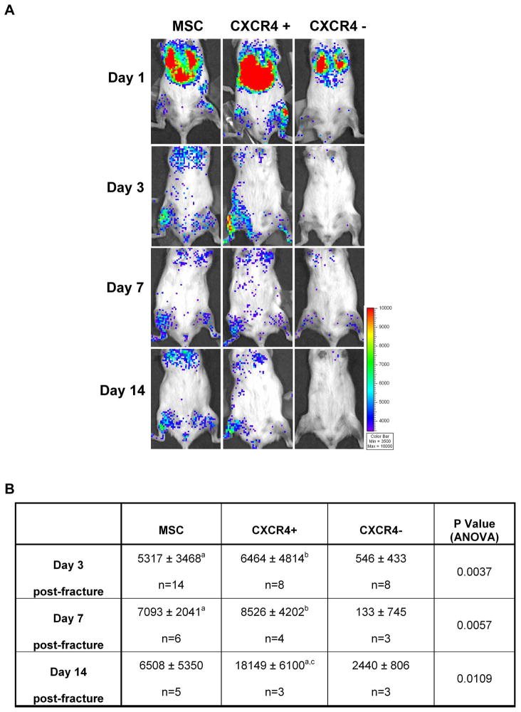
Figure 1. MSC migrate to the fracture site in a time- and CXCR4-dependent manner.
(A): BLI was performed at day 1, 3, 7 and 14 after fracture/transplant in mice with tibia fracture transplanted either with 106 MSC-β-Act-Luc (MSC) (left panel), MSC-β-Act-Luc-CXCR4+ (CXCR-4+) (middle panel) or MSC-β-Act-Luc-CXCR4-(CXCR-4−) (right panel). Graded color bar indicates BLI signal intensity expressed as photons/sec/cm2/sr. (B): BLI signal semi-quantitative analysis. Signal at the fracture tibia site ROI measured as photons/sec/cm2/sr, was normalized by subtracting the background signal found in an equal ROI in the contralateral unfractured tibia. a p<0.05 versus CXCR4-group; b p<0.01 versus CXCR4-group; c p<0.05 versus MSC by Tukey post-test. Abbreviations: MSC, mesenchymal stem cells.

