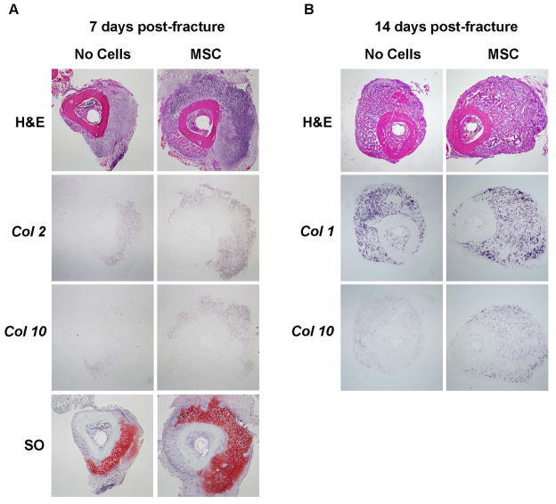
Figure 3. MSC transplant increases the cartilageneous and bone content of the callus.
(A): transversal sections of 7 days post-fracture calluses were subjected to H&E and Safranin O staining and in situ hybridization for Collagen-2 and Collagen-10. (B): 14 days post-fracture transversal sections were subjected to H&E staining and in situ hybridization for Collagen-1 and Collagen-10. The entire callus was sectioned (6 μm thick sections), the center of the callus was identified by the largest diameter of callus size by H&E staining and further analyses were performed within 20 sections from the center. Analyses were done in at least 5 sections for each probe or staining. Sections were obtained from at least 3 mice for each group. Abbreviations: H&E, hematoxylin & eosin; Col2, collagen 2; col1, collagen 10; SO, Safranin O; Col1, collagen 1; MSC, mesenchymal stem cells. 40X magnifications are presented.

