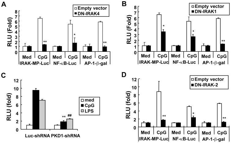
Figure 4. CpG DNA-mediated induction of Irak-m promoter activity is dependent on IRAK2 and PKD1 as well as IRAK4 and IRAK1.
Panels A, B, and D. RAW264.7 cells were transiently cotransfected with empty vector or plasmids encoding DN-IRAK4 (A), DN-IRAK1 (B), or DN-IRAK2 (D) and Irak-m-promoter-luciferase plus pRL-TK-luciferase reporters, NF-κB-luciferase plus pRL-TK-luciferase reporters, or AP-1-β-galactosidase reporter. Cells were stimulated with medium or CpG DNA (6 μg/ml). Luciferase activity in cell extracts was analyzed by the Dual-Luciferase Reporter Assay System and normalized using pRL-TK-luciferase activity in each sample. β-galactosidase activity in equal amounts of cell extracts was analyzed using the Galacto-Light Plus Reporter gene assay. Data are the mean relative light unit (fold induction from luciferase activity or β-galactosidase activity of the indicated reporter in the unstimulated cells) ± SD of triplicates. Significant differences from luciferase activity or β-galactosidase activity of the indicated reporter in the cells transfected with empty vector and stimulated with CpG DNA are indicated (* p<0.05; ** p<0.005). Panel C. Control luciferase-knockdown macrophages (Luc-shRNA) or Prkd1-knockdown macrophages (PKD-1shRNA) were cotransfected with Irak-m-promoter-luciferase and pRL-TK-luciferase. Transfected cells were treated with medium, CpG DNA (6 µg/ml), or LPS (50 ng/ml) for 36 hr. Luciferase activity in cell extracts was analyzed by the Dual-Luciferase Reporter Assay System and normalized using pRL-TK-luciferase activity in each sample. Data are the mean relative light unit (fold induction from luciferase activity of unstimulated cells) ± SD of triplicates. Significant differences from luciferase activity in Luc-shRNA cells stimulated with CpG DNA (** p<0.005) or LPS (## p<0.005) are indicated. All experiments were repeated at least three timeswith similar results.

