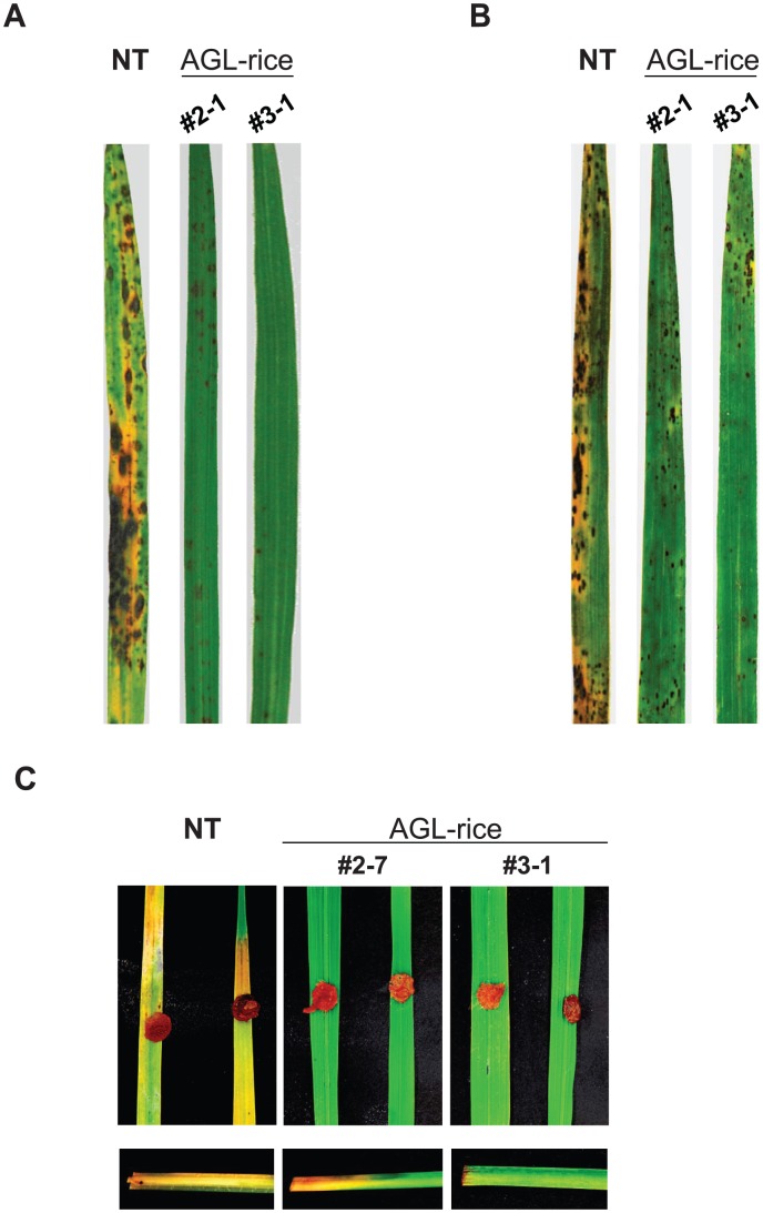
Figure 4. The fungal disease resistance of the transgenic rice expressing a bacterial α-1,3-glucanase (AGL-rice).
(A) Spray-inoculation assay of M. oryzae. Typical blast-disease lesions were observed on the non-transgenic (NT) leaves. In contrast, HR-like cell death was observed with the AGL-rice leaves (#2-1 and #3-1). The assay was repeated 20 times, and representative images are presented. The photos were taken at 6 dpi. (B) Spray-inoculation assay of Cochliobolus miyabeanus. Typical brown spot lesions were formed and coalesced on the NT rice leaves, while the symptoms were significantly decreased on the AGL-rice leaves. The assay was repeated 20 times, and representative images are presented. The photos were taken at 6 dpi. (C) Inoculation assay of Rhizoctonia solani. Rot was observed on the NT rice leaves, but not on the AGL-rice leaves. The rotted region on the culms was greatly reduced in the AGL-rice compared with the NT rice. Each assay was repeated 10 times, and representative images are presented. The photos were taken at 6 dpi.

