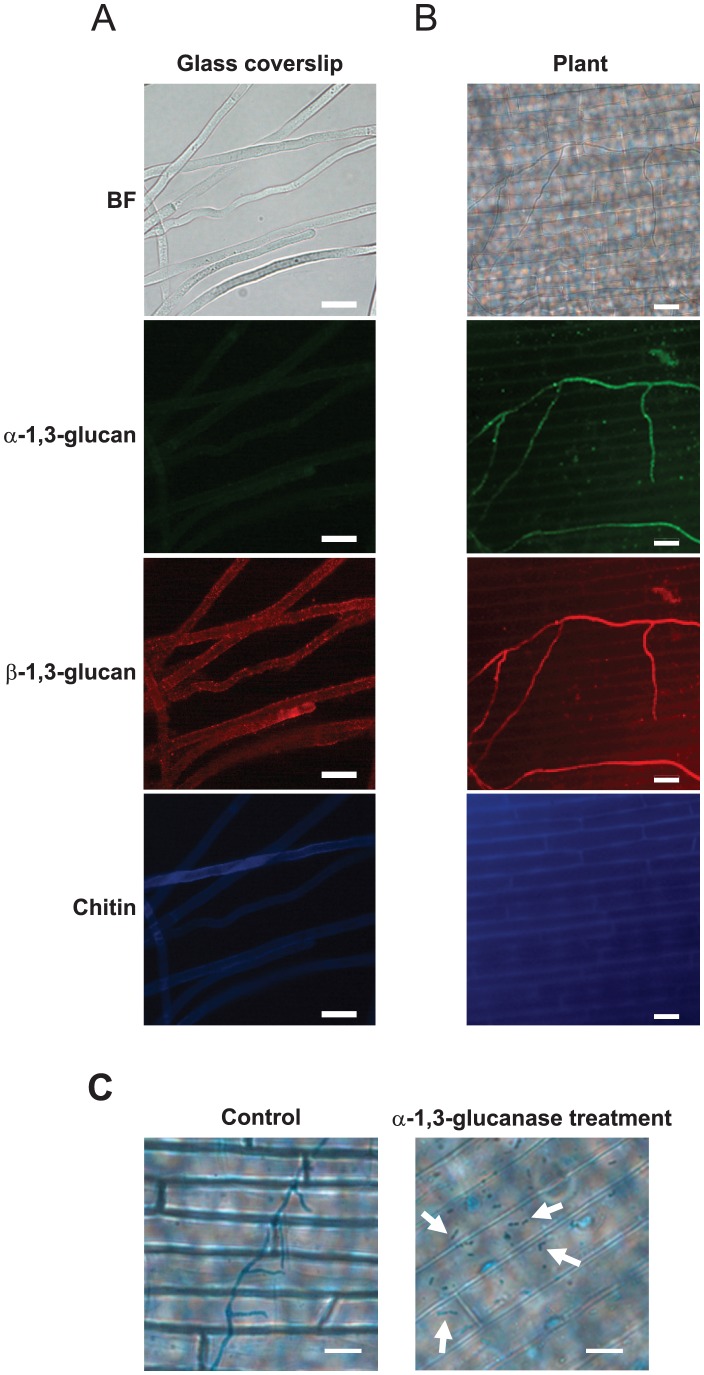
Figure 7. Detection of polysaccharides on the cell walls of R. solani hyphae.
α- and β-1,3-glucan were detected using fluorophore-labeled antibodies, and chitin was detected using a fluorophore-labeled lectin. (A) The detection of polysaccharides at the accessible surface of the cell wall of mycelia developed on glass coverslips. Representative images of 50 fungal cells from 10 independent experiments are presented. (B) The detection of polysaccharides at the accessible surface of the cell wall of infectious hyphae developed on rice sheath cells. The representative images of more than 150 mycelia from 15 independent experiments are presented. (C) The degradation of infectious hyphae after treatment with α-1,3-glucanase. The infectious hyphae developed on more than 15 rice sheaths from 5 rice plants were stained with lactophenol cotton blue and observed. BF: bright-field optics. Scale bar = 20 µm.

