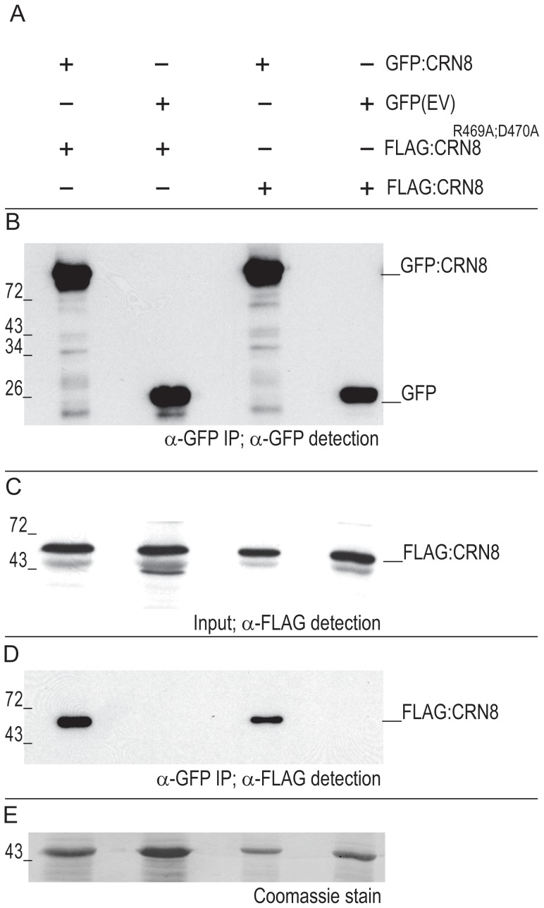
Figure 7. CRN8 forms a dimer.
(A) Overview of the different combinations of FLAG:CRN8, GFP:CRN8, FLAG:CRN8R469A;D470A, and GFP (indicated by +) proteins, co-expressed in N. benthamiana leaves. (B) Western blot of GFP:CRN8 and GFP protein from GFP immuno-purified protein samples probed with GFP antibody. (C) Western blot of FLAG:CRN8 and FLAG:CRN8R469A;D470A inputs for GFP co-immunopurification experiment probed with FLAG antibody. (D) Co-immunoprecipitation of FLAG:CRN8 and FLAG:CRN8 R469A;D470A in GFP immuno-purified protein samples on a Western blot probed with FLAG antibody. (E) Coomassie stain indicating equal loading of protein on Western blot in figure 7D.

