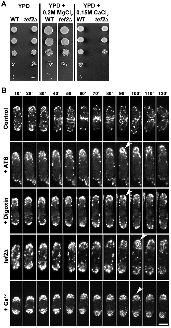
Figure 5. SpTef2 function and the F-actin cytoskeleton in S. pombe.
(A) Sensitivity of tef2Δ S. pombe cells towards Ca+2 in the growth medium. Serial dilutions of the wild-type or tef2Δ cells were inoculated under indicated growth conditions. (B) Morphology and dynamics of GFP-labelled F-actin cytoskeleton in wild-type S. pombe treated with ATS, digoxin or Ca+2. The tef2Δ strain was analyzed in parallel. Arrowheads show excess accumulation of F-actin patches and/or short, spooling cables at the cell end(s). The maximum projection images shown here represent the compressed z-stack sections. Bar equals 10 µm.

