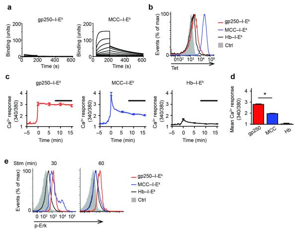
Figure 1.
gp250 ligand induces sustained Ca2+ signals in AND DP thymocytes. (a) Equilibrium binding of soluble AND TCR to biotinylated gp250–I-Ek and MCC–I-Ek ligand at various concentrations of soluble AND TCR. Sample SPR sensorgrams were used to determine binding affinity of soluble AND TCR to gp250–I-Ek (KD > 500 μM) or MCC–I-Ek (KD = 13.03 μM). n = 2. (b) Tetramerized biotinylated gp250–I-Ek or MCC–I-Ek staining of AND.Rag1−/−H-2d DP thymocytes. Hb–I-Ek and hCLIP/I-Ab tetramer (Ctrl) were used as negative controls. n = 3. (c) Fura-2 AM ratios for AND.Rag1−/−H-2d DP thymocytes stimulated with gp250–I-Ek, MCC–I-Ek, or Hb–I-Ek. Traces represent Fura-2 AM ratio mean ± s.e.m (n = 50) as a function of time. Curves for each cell (Supplementary Fig. 1) were adjusted to time 0 being when the Ca2+ influx started. Black bar indicates the time from 7.5 to 15 min used for the summary of mean ratios in (d) of 250 cells in five experiments. *P < 0.0001 by two-tailed Mann-Whitney test. (e) Erk phosphorylation of AND.Rag1−/−H-2d DP thymocytes induced by gp250–I-Ek, MCC– I-Ek, Hb–I-Ek at 30 min and 60 min of stimulation time.

