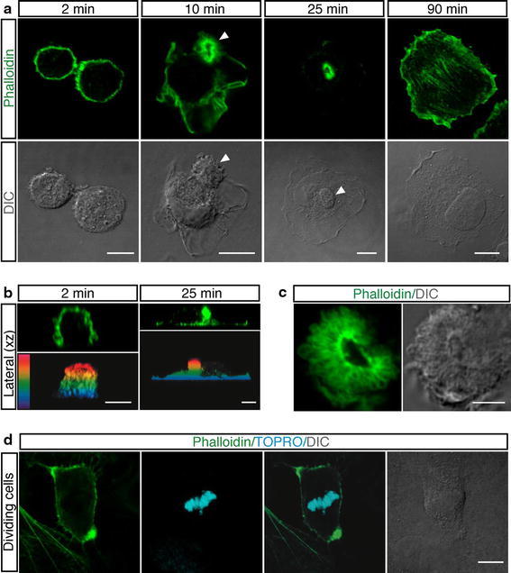
Fig. 1.

Spreading of round endothelial cells is characterized by the formation of an actin-rich domain in the cellular apical membrane. a HUVEC were trypsinized and resuspended in complete medium. To examine different phases of spreading, cells were allowed to settle on vitronectin-coated coverslips, fixed at the indicated times after plating and stained with phalloidin (green). Arrowheads indicate the actin-rich domain. Scale bars represent 8 μm. b Periodic z-series of HUVEC treated as in a were acquired at 2 and 25 min after plating to generate vertical sections (lateral xz). The same images are shown as color-coded projections such that the basal cell surface, which adheres to the substrate is violet and the apical cell surface is red. Scale bars 6 μm. c Details of an actin-rich domain formed on the apical surface of spreading HUVEC treated as in a. Scale bar 4 μm. d HUVEC were plated on coverslips coated with vitronectin. After 2 h, the culture medium was removed, cells washed with PBS and fresh medium added. 20 h after medium removal, samples were stained for F-actin (green) and DNA (blue). Mitotic cells display actin-rich domains located at the cellular edges. Scale bar is 8 μm. DIC images are shown
