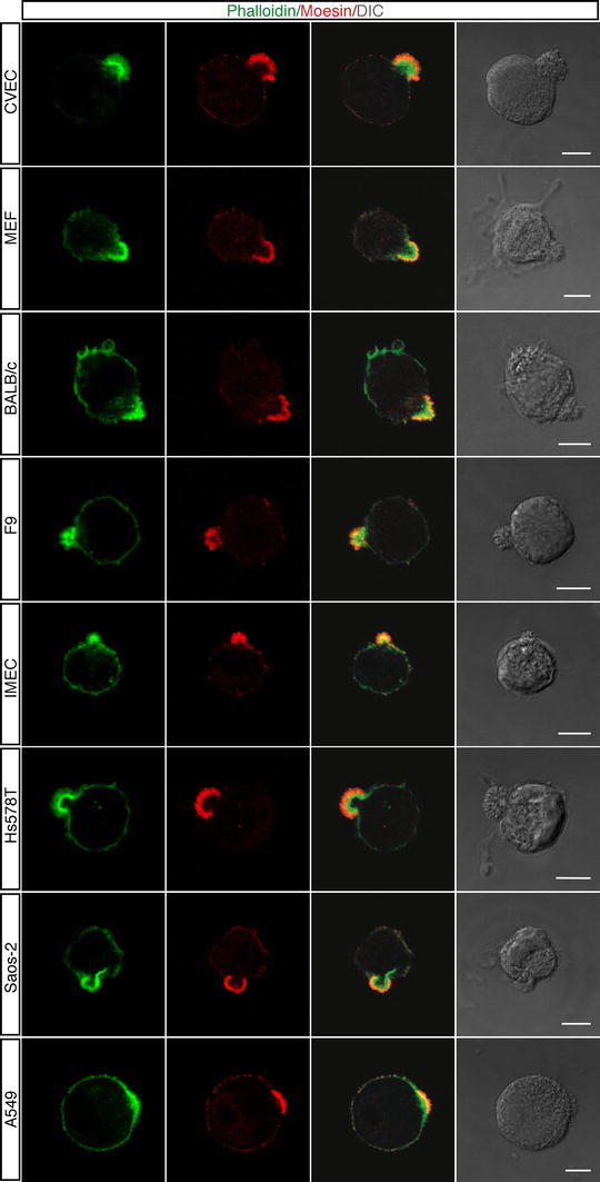
Fig. 2.

Different cell types display an apical domain enriched in F-actin and Moesin during adhesion to ECM. Cells were grown using standard conditions, detached from the plate, resuspended in complete medium and plated on vitronectin. Cells were analyzed during the initial phase of spreading using phalloidin (green) and anti-Moesin antibodies (red). DIC images of stained cells are shown. Scale bars represent 7 μm
