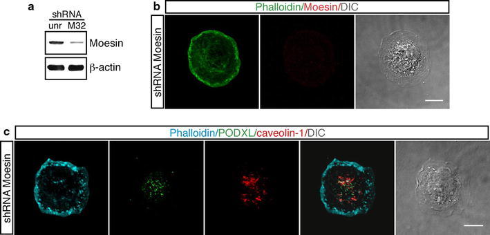
Fig. 6.

Effects of Moesin silencing in endothelial cells during the early phases of adhesion. a HUVEC were infected with a lentiviral vector expressing unrelated (unr) or Moesin (clone M32) shRNA. Cell extracts from HUVEC expressing shRNA were analyzed by western blotting using anti-Moesin and anti-β-actin antibodies. Cells infected with lentiviruses expressing Moesin shRNA show a reduced protein expression with respect to cells infected with a lentivirus expressing an unrelated shRNA. b Endothelial cells infected as in a were detached from the plate, seeded on vitronectin-coated coverslips, and fixed at different times after plating. Cells were stained for F-actin (green) and Moesin (red). Moesin-silenced cells do not form apical buds during spreading. Scale bar is 10 μm (c). Cells treated as in b were stained for F-actin (blue), PODXL (green) and caveolin-1 (red). PODXL and caveolin-1 are randomly distributed into the cell. Scale bar represents 8 μm. Representative adhering cells fixed after 8 min from plating are shown. DIC images of stained cells are shown
