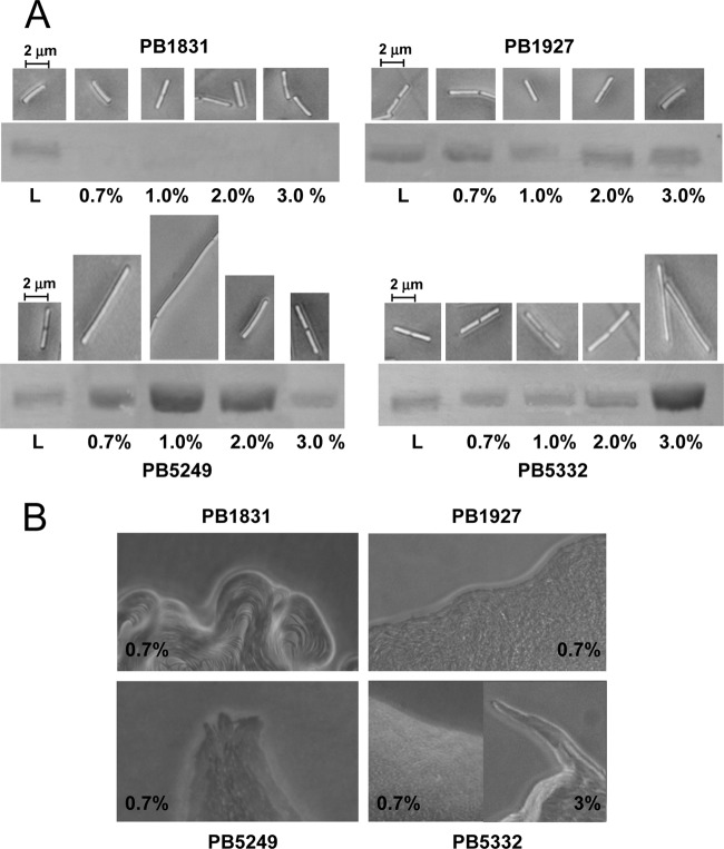
Fig 3.
Analysis of swarming features of B. subtilis strains. (A) Cell length (images obtained by phase-contrast microscopy) and surface flagellin (SDS-PAGE of extracellular flagellin) of strains grown on Tr plates solidified with different percentages of agar. L, liquid cultures. (B) Colony edges of B. subtilis strains grown on Tr medium containing 0.7% or 3% agar (magnification, ×200).

