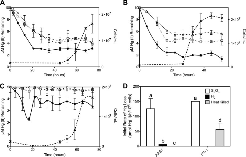
Fig 2.
203Hg(II) remaining in media during growth of strains AAS1 (A and C) and R1-1 (B) using S2O32− (A and B) and H2 (C) as electron donors. Cell density (×) is shown, as is loss of Hg(II) (in μM) from growing cultures (●), heat-killed controls (□), and uninoculated controls (△). (D) Initial rates of Hg(II) loss [μmol Hg(II)/h/106 cells] calculated for strains AAS1 and R1-1 in cultures grown on S2O32− or H2 and for heat-killed controls. Different letters indicate statistical significance (P < 0.05). All data represent the mean Hg(II) loss of triplicate growing cultures ± 1 standard deviation (SD) after subtracting loss rates of uninoculated controls.

