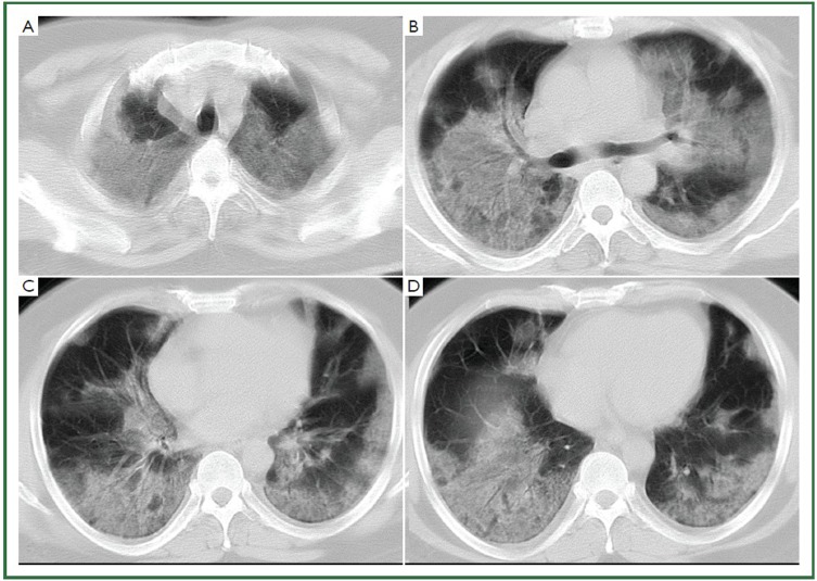
Figure 2.
44-year-old man with H1N1 viral pneumonia in critical condition. No history of chronic pulmonary disease. Chest CT was obtained 6 days after the onset of the influenza symptoms. Bilateral and widespread ground-glass opacities in all six lung zones are demonstrated (A-D). There is bilateral mild pleural effusion. The chest lesion score is 19.

