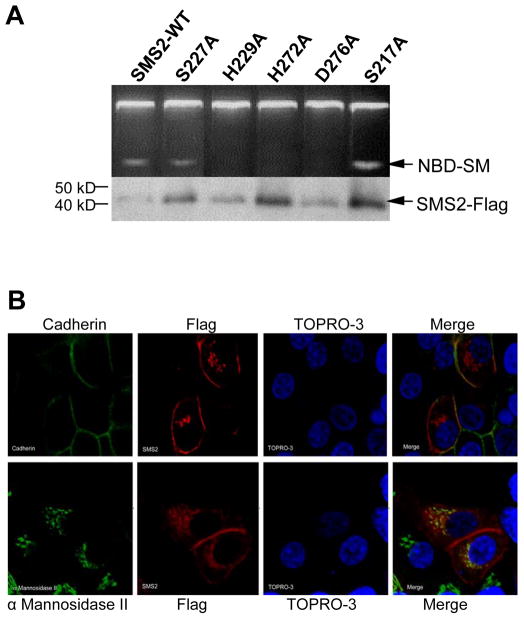
Fig. 3.
Point mutation of individual conserved residues within D3 and D4 abolishes SMS2 activity with the exception of S227. (A) SMS activity was performed on wild type (WT) or mutant SMS1 following immunoprecipitation from Hela cells transiently expressing the respective enzyme. The protein levels of each enzyme is shown below. (B) Wild type SMS2 localizes to both the plasma membrane and Golgi. Both cadherin (top) and α Mannosidase II (bottom) are depicted in green, while SMS2-Flag is depicted in red. TOPRO-3 is in blue. (C) Each SMS1 mutant (S2227A, H229A, H272A, H276A, S217A) exhibits a WT localization pattern. Cadherin is depicted in green. Flag-tagged SMS1 and mutants are depicted in red. TOPRO-3 is in blue.


