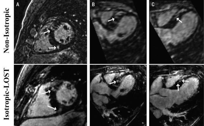
Figure 4:
Reformatted 3D LV LGE MR images (5.2/2.6; section thickness, 4 mm [nonisotropic] and 1.5 mm [isotropic]; matrix, 376 × 235; field of view, 320 × 320 cm2) in, A, short-axis, B, four-chamber, and, C, three-chamber views obtained with nonisotropic and isotropic resolution in 46-year-old man with hypertrophic cardiomyopathy. Isotropic resolution allows superior visualization of scar morphology in three-chamber view (C), where two discrete areas of LGE are visualized; only one area of enhancement is seen with nonisotropic imaging. Arrows = regions of enhancement.

