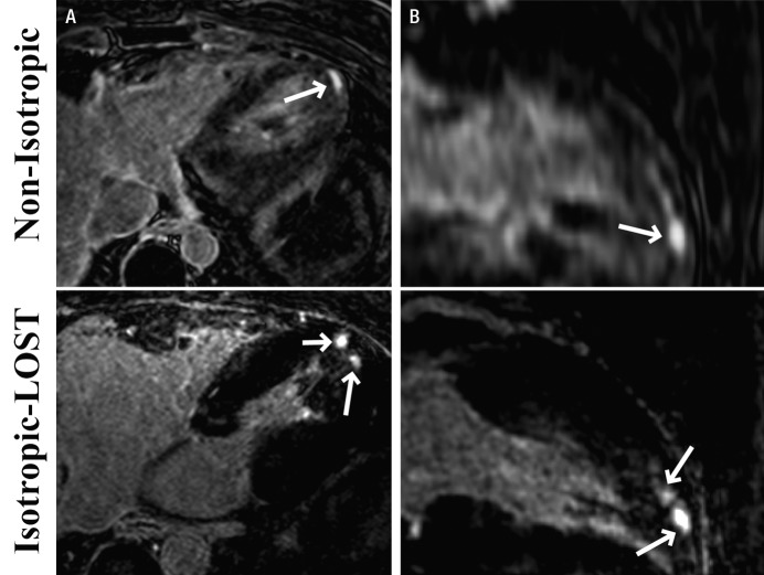
Figure 6:
A, Axial and, B, reformatted long-axis 3D LV LGE MR images (5.2/2.6; section thickness, 4 mm [nonisotropic] and 1.2 mm [isotropic]; matrix, 536 × 319; field of view, 320 × 320 cm2) obtained with nonisotropic and LOST-accelerated isotropic spatial resolution in 32-year-old man. Scar morphology (arrows) is visualized in greater detail on isotropic LOST images.

