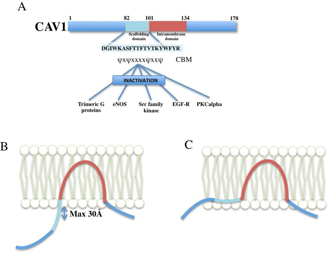
Figure 1.
The caveolin signalling hypothesis. (A) Schematic of the caveolin signalling hypothesis as originally proposed (Okamoto et al., 1998), with some key interacting partners highlighted. The sequence of the caveolin-1 scaffolding domain (CSD) and the consensus caveolin-binding motif (CBM) are shown. (B) and (C); Two models for caveolin association with the membrane bilayer. In model (B) the CSD is exposed and shown in an extended conformation allowing interactions with signaling proteins. However, note that the middle of the CSD is still very close to the membrane, even assuming a completely extended polypeptide conformation perpendicular to the bilayer. Model (C), in which the CSD forms part of an amphipathic cholesterol-binding in-plane helix, is an alternative model supported by a number of studies (Kirkham et al., 2008).

