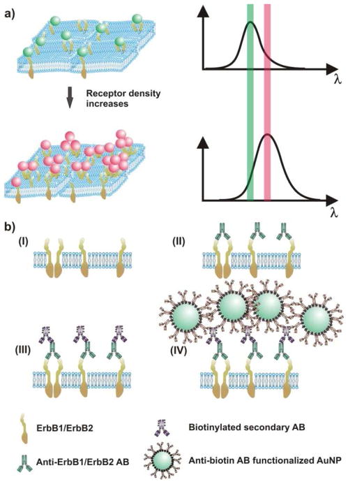
Figure 1. The spectral response of NP immunolabels depends on the NP density and clustering on the cell surface.
a) As the average interparticle separation between the NP decreases (left) the area-averaged plasmon spectrum red-shifts and increase in scattering intensity (right). b) Labeling strategy for cell-surface ErbB1 and ErbB2: (I) ErbB receptors embedded in the plasma membrane are (II) labeled with primary antibodies (ABs). (III) Biotinylated secondary ABs are tethered to the primary antibodies before, in the final step (IV), anti-biotin AB-functionalized NPs are targeted to biotin binding sites.

