Fig. 1.
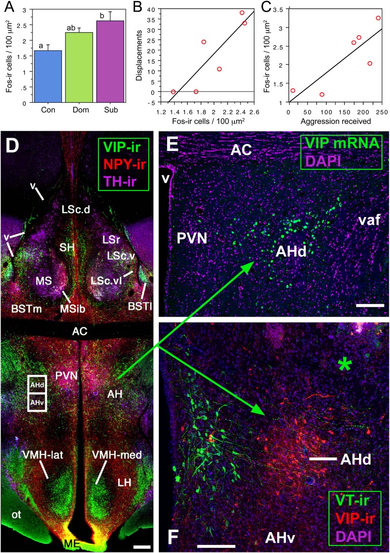
Histochemically distinct components of the AH exhibit differential responses to agonistic encounters. (A) Fos-ir cell numbers in the AH are significantly increased in subordinate (Sub) but not dominant (Dom) male waxbills relative to control subjects (Con); n = 6 per group. Different letters above the bars denote significant pairwise differences (P < 0.05). (B and C) Fos activation in the dorsal AH (AHd) of dominant birds correlates positively with the number of aggressive displacements given (r2 = 0.752, P = 0.02; B), whereas Fos activation in the ventral AH (AHv) of subordinate birds correlates positively with aggression received (r2 = 0.704, P = 0.04; C). (D–F) VIP elements in the septohypothalamic region of the zebra finch (D and E) and violet-eared waxbill (F; asterisk shows the injector tract in a scrambled oligonucleotide subject). The dorsal AH region is defined by VIP mRNA and peptide (E and F). (Scale bars, 200 μm in D and 100 μm in E and F.) AC, anterior commissure; AH, anterior hypothalamus; BSTl; lateral bed nucleus of the stria terminalis; BSTm, medial BST; LS, lateral septum (LSr. rostral LS division; LSc., caudal LS division: d, dorsal; v, ventral; vl, ventrolateral); ME, median eminence; MS, medial septum; MSib, internal band of the MS; ot, optic tract; PVN; paraventricular nucleus; SH, septohippocampal septum; v, ventricle; vaf, ventral amygdalofugal tract. D is modified from ref. 17.

