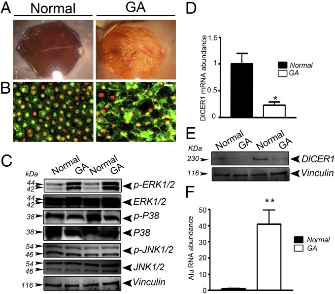
Fig. 1.
DICER1 deficit in human GA RPE is accompanied by an increase in Alu RNA and ERK1/2 phosphorylation. (A) Fundus photographs and (B) flat mounts stained for ZO-1 (red) show cell degeneration and atrophy in RPE from a human GA eye compared with a normal eye. (Scale bar, 20 μm.) (C) ERK1/2 phosphorylation, assessed by Western blotting, is increased in human GA RPE compared with control RPE; however, p-p38 MAPK and p-JNK1/2 remained unchanged between the two groups. DICER1 quantification, assessed by quantitative PCR (D) and Western blotting (E), is lower in human GA RPE compared with control RPE (n = 5 independent experiments, *P = 0.012 by Student’s t test). (F) Alu RNA abundance is increased in human GA RPE compared with control RPE (n = 5 independent experiments, **P < 0.0001 by Student’s t test). Images are representative of three independent experiments (C and E).

