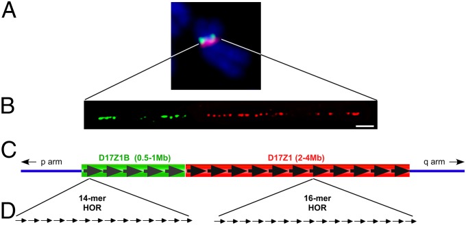
Fig. 1.
Genomic organization of HSA17 centromere. (A) HSA17 contains two HOR alpha-satellite arrays, D17Z1 (red) and D17Z1-B (green), that can be identified by high-stringency FISH. D17Z1-B is located on the short (p) arm side of the centromere. (B) Stretched chromatin fibers hybridized with D17Z1 (red) or D17Z1-B (green) FISH probes confirm that the two arrays are juxtaposed. (C and D) D17Z1 primarily contains reiterated HOR units composed of 16 individual 171-bp alpha-satellite monomers that are tandemly arranged. Overall, the array is estimated to cover 2–4 Mb. The HOR of D17Z1-B contains 14 monomers, and the entire array is estimated to be one-fourth the size of D17Z1. (Scale bar: 10 microns.)

