Fig. 2.
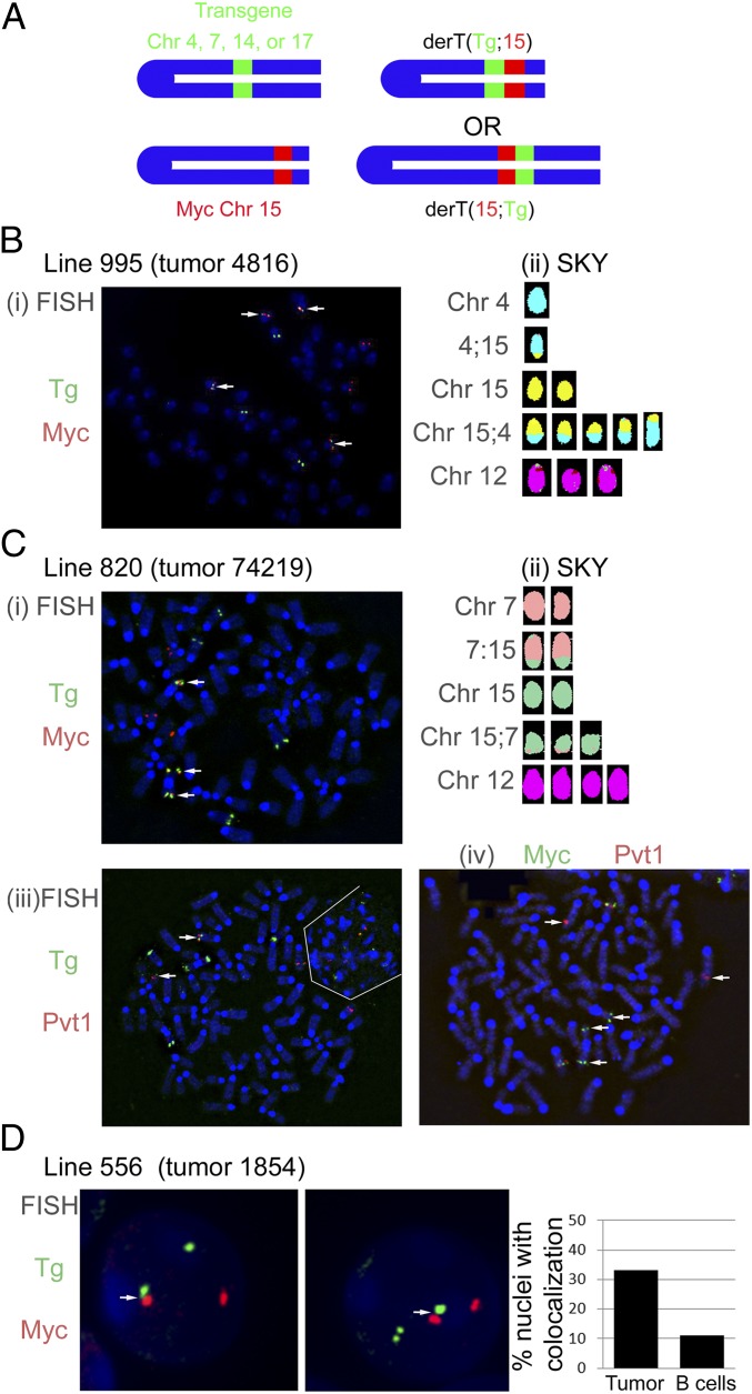
Plasmacytomas harbor translocations to the Igh transgene. (A) Colocalization of the Igh transgene with Myc or Pvt1. Colocalization of BACs spanning the Igh transgene insertion site on Chr 4 (line 995), 7 (line 820), or 17 (line 556) and either Myc or Pvt1 reveal translocations involving the two loci. White arrows note separation of probes (Civ) or colocalization (C i–iii). (B) Two-color FISH (i) and SKY (ii) on metaphase spreads from tumor no. 4816, line 995. The color coding for Chr 15 for the SKY analysis of these metaphases was changed to yellow to better contrast with the aqua color coding for Chr 4. (C) Two-color FISH and SKY analysis of tumor no. 74219, line 820. In Ci, the Igh transgene is shown to colocalize with Myc. In Ciii, the Igh transgene is shown to colocalize with Pvt1. The white lines delimit a second, interphase cell that lies next to the metaphase. In Civ, the Myc and Pvt1 probes are rearranged onto different chromosomes. (D) Interphase two-color FISH analysis of tumor no. 1854, line 556 (two representative cells shown). Myc and the Igh transgene were colocalized in 33% of the interphases from tumor no. 1854 ascites, and in 11% of the interphases from normal B-cell controls (P < 0.001, Fisher’s exact test).

