Fig. 1.
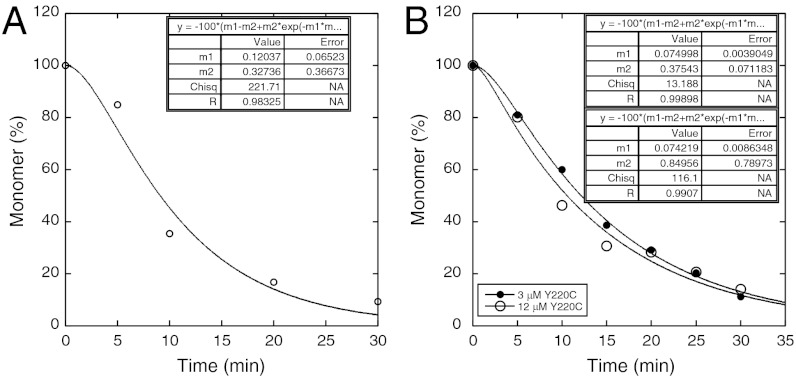
Soluble p53 monomer remaining in supernatant during aggregation of Y220C at 37 °C. (A) Monomer peak from gel filtration assay. (B) SDS-PAGE stained by Sypro Orange for aggregation initiated from 3 μM (Left) and 12 μM (Right) Y220C. Monomer depletion curves obtained by (C), gel filtration assay, and (D), SDS-PAGE assay fitted to Eq. 1.

