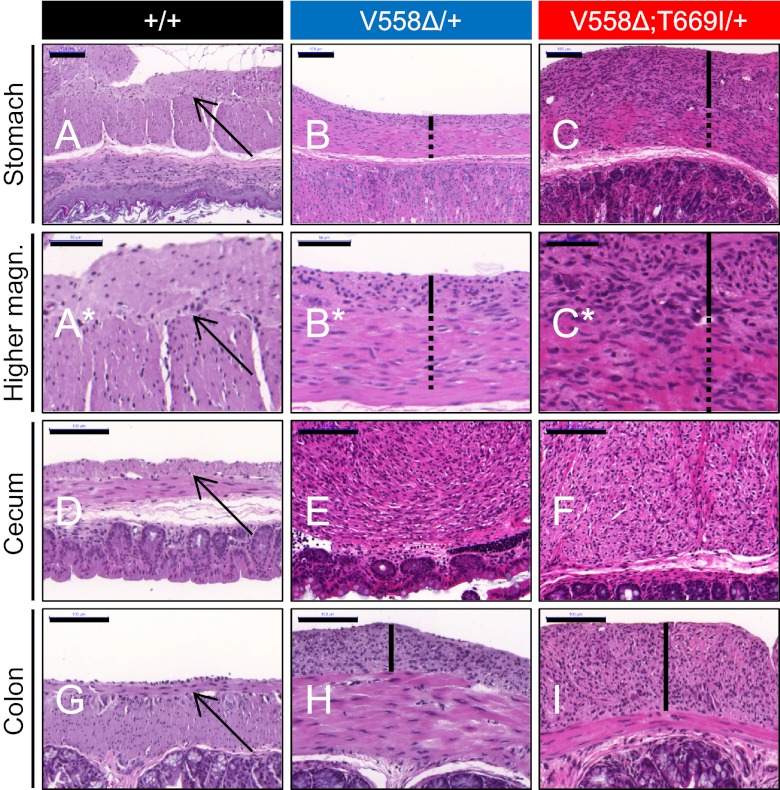
Fig. 2.
Cecal GIST and pronounced gastric and colonic ICC hyperplasia in KitV558Δ;T669I/+ mice. Cross-sections of stomach (A–C; higher magnification is shown in A*–C*), cecum (D–F), and colon (G–I) of 3- to 4-mo-old wild-type, KitV558Δ/+, and KitV558Δ;T669I/+ mice. Arrows indicate normal thin layer of myenteric ICC in wild-type samples. ICC hyperplasia in stomach and colon samples is indicated by black bars. Note the extensive hyperplasia involving the circular muscle layer (dotted lines) in the stomach of KitV558Δ;T669I/+ mice. Photographs show representative H&E staining; n ≥ 3 each. (Scale bars: 50 μm in A*–C*; 100 μm in A–I.)

