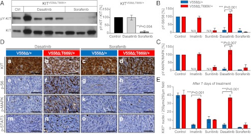
Fig. 4.
Resistance to dasatinib and sensitivity to sorafenib treatment in KitV558∆;T669I/+ mice. (A–C) Tumor lysates from individual KitV558Δ/+ and KitV558∆;T669I/+ animals treated with vehicle, imatinib, sunitinib, dasatinib, or sorafenib were subjected to Western blotting to detect the abundance of phosphorylated and total KIT, S6, and MAPK proteins. Representative blots for phospho-Y719-KIT and KIT are shown. The ratio of phosphorylated to total protein was quantified by densitometry. n = 3–4 animals per treatment group; error bars indicate means ± SD. (D) Representative results after short-term treatment with dasatinib and sorafenib; IHC on GIST sections with antibodies as specified in Fig. 3D. (E) Cell proliferation in GIST of KitV558Δ;T669I/+ mice is unaffected by long-term treatment with imatinib and dasatinib. Sunitinib and sorafenib overcome resistance and attenuate cell proliferation. Ki67+ nuclei per 250 × 250 μm field; n ≥ 3 each; error bars indicate means ± SD.

