Fig. 5.
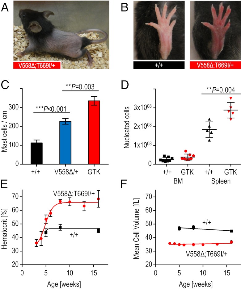
Increased mast cell and red blood cell numbers in KitV558∆;T669I/+ mice. (A) Representative photograph of alopecia of truncal hair in 4-wk-old KitV558Δ;T669I/+ mice. (B) Representative photographs of wild-type hind-paw in 3-mo-old wild-type mice and “red paw” phenotype in 3-mo-old KitV558Δ;T669I/+ mice. (C) Increased mast cell numbers in dorsal skin of KitV558Δ;T669I/+ (GTK) mice in comparison with wild-type and KitV558Δ/+ mice (n = 3). (D) BM cellularity of KitV558Δ;T669I/+ mice measured as total nucleated cells per bone is similar to that in wild-type mice. Spleen cellularity is significantly increased in the mutants compared with wild-type mice. n = 6. (E) Time course of hematocrit and (F) MCV showing development of erythrocytosis and microcytosis phenotype in KitV558Δ;T669I/+ mice (red curves) in and wild-type mice (black curves). n = 5–17. Error bars indicate means ± SD.

