Fig. 3.
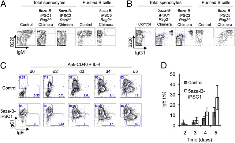
The 5aza-B-iPSCs can form all-IgG1 chimeric mice in Rag2−/− hosts, which can support further CSR to IgE. (A) FACS plots of splenocytes from 5aza-B-iPSC–complemented Rag2−/− chimeras stained for surface expression of the pan–B-cell marker B220 and IgM. For comparison, splenocytes from reprogrammable mice indicated as control were analyzed in parallel. Cells expressing both B220 and IgM are demarcated within FACS plots by a box in the upper right-hand corner. (B) FACS plots of splenocytes from control mice and 5aza-B-iPSC–complemented Rag2−/− chimeras stained for surface expression of B220 and IgG1. Cells expressing both B220 and IgG1 are shown by a box in the upper right-hand corner. (C) FACS analysis of day 0, 2, 3, 4, and 5 anti-CD40/IL-4–stimulated B cells from reprogrammable mice (control) or 5aza-B-iPSC–derived B cells stained for cytoplasmic IgG1 and IgE expression as described in the text. Gates within the plots demarcate IgG1+ and IgE+ B cells in the upper left and lower right portion of box, respectively. Numbers indicate percentage of cells within the gates. (D) Statistical analysis of three independent FACS analysis experiments for IgE switching of control and 5aza-B-iPSC–derived B cells activated with anti-CD40 plus IL-4 for the indicated time points. Shown are mean values ± SD of three independent experiments involving Rag2−/− iPSC chimeras derived from 5aza-B-iPSC#2.

