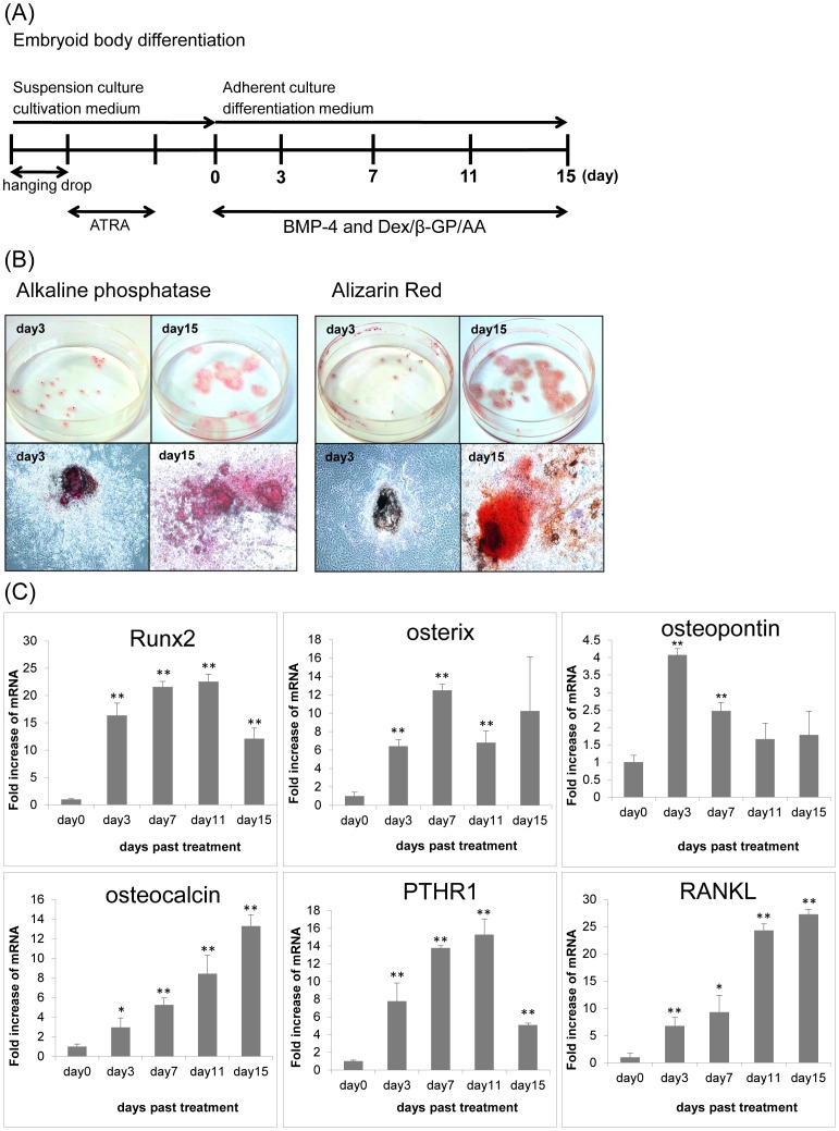
Figure 1. Osteoblast differentiation induced by BMP-4 in iPS cells.
(A) Schematic representation of the osteoblast differentiation protocol for iPS cells by osteogenic cocktail. (B) Histochemical staining for ALP activity and with alizarin red in differentiated iPS cells on day 3 and day 15. EBs were grown for 15 days in the presence of Dex/h-GP/AA with BMP-4 and then stained (ALP and alizarin red). (C) Time course of osteoblast marker expression in osteoblastic differentiated iPS cells. Quantitative RT-PCR for osteoblast markers was carried out. Bars represent means ± SD of 3 separate wells.

