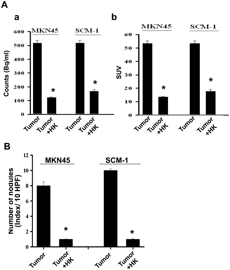
Figure 3. Quantification of honokiol-inhibited peritoneal metastasis of gastric tumor cells.
Mice were inoculated with human gastric cancer cells (MKN45 and SCM-1). [18F]-FDG-PET/CT imaging was performed and analyzed. (A) Quantifications of estimated radioactivity (Bq/ml, a) and specific uptake values (SUV, b) were calculated. SUV is used as an index to determine if a hotspot is significant. (B) Photomacrographs of metastatic peritoneal nodules are shown. All data are presented as mean ± SEM (n = 6–8). *p<0.05 as compared with control.

