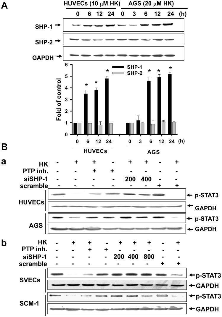
Figure 10. Honokiol enhanced phosphatase SHP-1 protein expression and the interaction with STAT-3 in gastric cancer cells and HUVECs.
Cells were treated with honokiol (10 and 20 µM) for various time courses as indicated. (A) SHP-1 and SHP-2 protein levels were detected by Western blot analysis in cells with or without honokiol treatment. (B) The phosphorylation of STAT-3 in gastric cancer cells (AGS and SCM-1) and endothelial cells (HUVECs and SVECs) with or without honokiol (10 µM in HUVECs, SVECs, and SCM-1; 20 µM in AGS) treatment for 24 h in the presence or absence of a phosphatase inhibitor (PTP inhibitor II, 20 µM) or SHP-1 siRNA transfection was detected. In A, data are presented as mean ± SEM of five independent experiments. *p<0.05 as compared with control. In B, the results shown are representative of at least four independent experiments.

