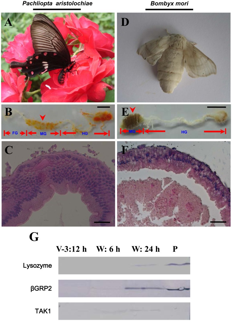
Figure 1. Different morphology of silkworm moth midgut compared with that of a butterfly.
(A–C) Morphology of a butterfly (Pachliopta aristolochiae) and its adult midgut; (D–F) Morphology of a silkworm moth (Bombyx mori) and its adult midgut. The silkworm moth’s foregut (FG) is not easily dissected with the midgut. In (B and E), the arrow-indicated part of midgut (MG) was sampled for histological study as shown in (C and F). The butterfly (A) ingests nectar and the midgut has many layers of cells (C) and appears in good condition. However, the midgut of silkworm moth is full of yellow body debris that cannot be excreted (E) and appears in weak condition due to one layer of cells (F). The silkworm moth (D) does not ingest anything and dies after egg-laying. FG: foregut; MG: midgut; HG: hindgut. (G) Three immunity-related proteins were significant expressed in the midgut during the wandering stage. Some proteins, such as lysozyme, βGRP2 (antibody against M. sexta βGRP2; 31% similarity to B. mori βGRP2), and TAK1 (antibody against mouse TAK1; 70% similarity to B. mori TAK1) were detected in the midgut during the wandering stage. Plasma (P) was from larvae injected with E. coli. For each lane, approximately 10 µg cell lysate was loaded. Bars: (B and E) 4 mm; (C and F): 50 µm.

