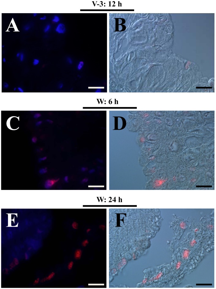
Figure 10. Apoptotic cells in the midgut. Midguts from larvae during the feeding (V-3: 12 h) and wandering (W: 6 h and W: 24 h) stages were sampled.
Very few TUNEL-positive (red) cells were found in the midgut during the feeding stage (A, B). However, many cells were undergoing apoptosis in the midgut during the wandering stage (C, D, E, F). Before pupation (W: 24 h), old midguts were observed to slough off from the outer layer of basement membrane. DAPI was used for nuclei counter-staining. All images were merged from pictures taken using red and blue filters or using red and DIC (Nomarski) filters. Bars: 20 µm.

