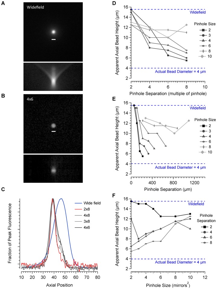
Figure 3. Characterisation of axial resolution.
A. Maximum projection of wide-field images of a 4 µm bead taken over an axial distance of 120 µm at 0.5 µm intervals. Below is an axial projection from a frontal plane. B. Images taken of the same bead using a 4×6 pinhole configuration and processed in the same way. Axial projections were made for a range of different pinhole configurations and line profiles drawn through the centre of the bead. C. Axial line profiles comparing 4 different pinhole configurations with images taken in wide-field are shown. Data were fitted with a Gaussian curve to estimate the axial height of the bead. Figures D–F illustrate the relationship between axial resolution, pinhole size and pinhole separation.

