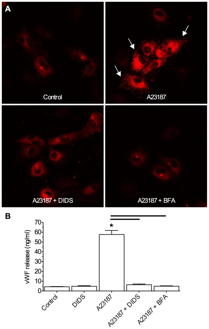
Figure 8. DIDS inhibits stimulus induced vWF release from normoxic HUVECs.
The Ca2+ ionophore A23187 induces vWF extrusion from HUVECs in a non-pathological model of vesicular release. DIDS abolishes stimulus-evoked vWF release. (A) Confocal Z-stack projection fluorescent images of vWF localization (red) in HUVECs treated as indicated. Arrows indicate vWF released extracellularly. (B) Summary of supernatant vWF expression measured by ELISA. Data are mean ±SEM from 3 separate 15-min experiments. Asterisks (*) indicate significant difference from normoxic controls; black bars indicate significance between connected treatments (p<0.05). Treatments as per Fig. 1 caption, and 10 µl A23187, 1 µg/ml brefeldin A (BFA).

