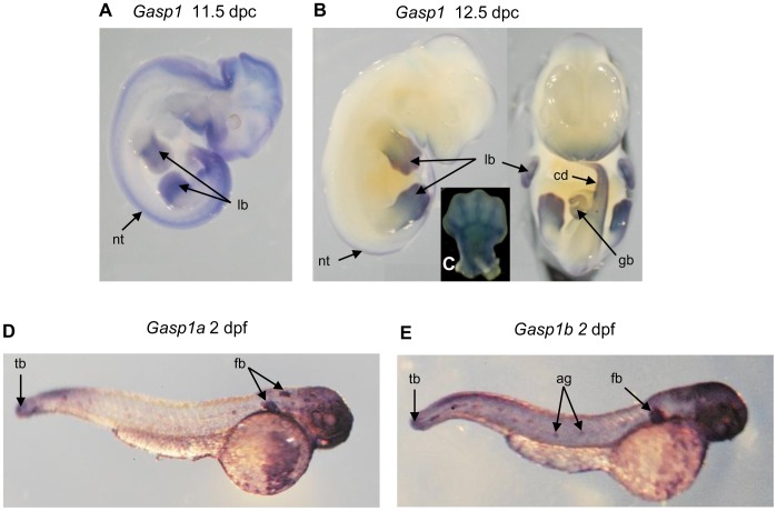
Figure 5. Gasp1 expression patterns.
A and B: in situ hybridization with a Gasp1 probe on mouse embryos at 11.5 dpc and 12.5 dpc reveals expression in neural tube (nt), limb bud (lb), genital bud (gb) and caudal part of the embryo (cd). Enlargement of the limb bud (C) shows an expression in the precartilaginous condensations that give rise to fingers. D and E: in situ hybridization with Gasp1 probes on zebrafish embryos at 2 dpf showing expression patterns in tail bud (tb), fin buds (fb) and angioblasts (ag).

