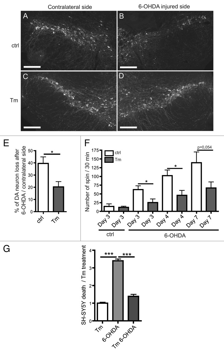
Figure 2. Tm is protective in the 6-OHDA mouse Parkinson disease model. (A–D) Sections of the substantia nigra (SN) obtained 4 d after 6-OHDA treatment with or without Tm pre-treatment (0.1 mg/kg). DA neurons are visualized by immunostaining for tyrosine hydroxylase (TH). (E) Quantification of DA neuron loss after 6-OHDA injection normalized to the contralateral side. (F) Rotational behavior of mice after 6-OHDA injection. The graph shows the number of unilateral turns made by mice on days 3, 4 and 7 after the 6-OHDA injection with or without Tm (n = 6–7). (G) In vitro experiments on SH-SY5Y to assess the cell viability after Tm and 6-OHDA treatments. Cell viability was evaluated using trypan blue after Tm and 6-OHDA treatments. Quantification of cell death is normalized to Tm treatment. *p ≤ 0.05, ***p < 0.001 in Student’s t-test. Scale bar: 200 µm.

An official website of the United States government
Here's how you know
Official websites use .gov
A
.gov website belongs to an official
government organization in the United States.
Secure .gov websites use HTTPS
A lock (
) or https:// means you've safely
connected to the .gov website. Share sensitive
information only on official, secure websites.
