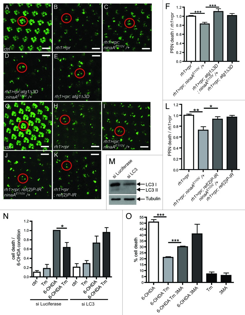
Figure 4. Autophagy is required for ER-mediated neuroprotection. (A–E and G–K) Visualization of PRN viability in 16 h-old living flies expressing rh1 > GFP. (A–E) Visualization of PRN in retina overexpressing rpr (rh1 > rpr) and mutant for ninaAE110V/+ and atg1Δ3D. (F) Quantification of PRN loss in the various mutants (B–E) relative to rh1 > rpr (n = 10). (G–K) Visualization of PRN in retina overexpressing rpr (rh1 > rpr), ref(2)P-IR and mutant for ninaAE110V/+. (L) Quantification of PRN loss in the various mutants (H–K) relative to rh1 > rpr (n = 10). (M) western blotting showing LC3I/II levels after siRNA against LC3 in SH-SY5Y cell compared with control (siRNA luciferase). (N) SH-SY5Y cell viability was assessed by trypan blue exclusion after treatments with Tm, 6-OHDA and siRNA against LC3 or luciferase as control (n = 3). (O) SH-SY5Y cell viability was assessed by trypan blue exclusion after Tm and 6-OHDA treatments. 3-MA treatment is used to block autophagy. *p ≤ 0.05, **p < 0.01, ***p < 0.001 in Student’s t-test. Scale bar: 10 µm. The abbreviations used: rh1-gal4; UAS-GFP (rh1 > GFP), rh1-gal4;UAS-rpr (rh1 > rpr).

An official website of the United States government
Here's how you know
Official websites use .gov
A
.gov website belongs to an official
government organization in the United States.
Secure .gov websites use HTTPS
A lock (
) or https:// means you've safely
connected to the .gov website. Share sensitive
information only on official, secure websites.
