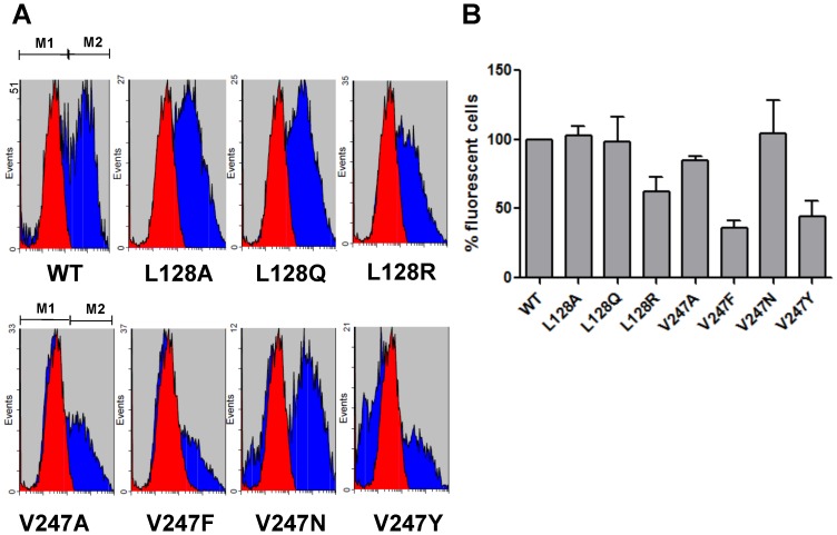
Figure 2. FACS analysis of CXCR1 WT and mutants.
HEK 293 cells were transiently transfected with CXCR1 or its mutants. Cells were incubated with FITC-conjugated mouse anti-human CD181 (CXCR1) antibody. Specificity of signal was confirmed by staining the cells with mouse IgG1 isotype control. (A) Transfected HEK 293 cell population stained positive for this anti-CXCR1 antibody (M2 region). (B) Fluorescence of positively stained cells was quantified by FACS (± SEM, with CXCR1 WT set to 100%).

