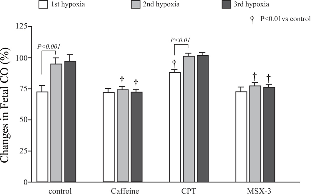
Figure 5. Mean percentage changes in fetal cardiac output following the onset of hypoxia.
The mean percentage change of the fetal cardiac output after the first hypoxia in the CPT group was higher than that in the control group. Control and CPT groups showed a similar trend of changes in fetal CO, while Caffeine and adenosine A2A antagonist, MSX-3, treated group did not recover the fetal CO to the baseline levels following 2nd and 3rd hypoxia exposure.

