Figure 4.
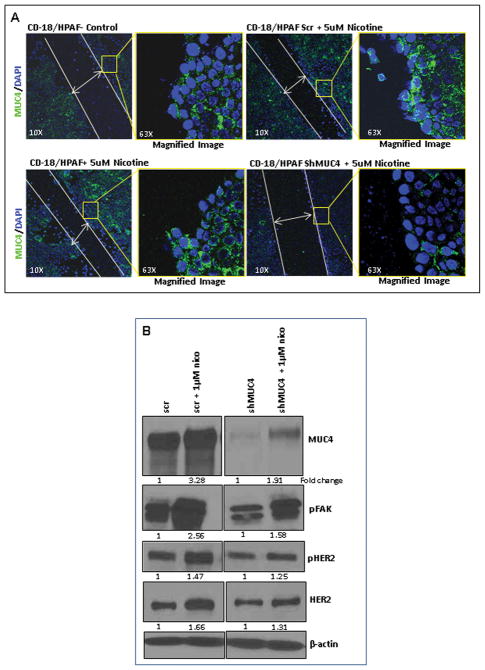
Nicotine-mediated effects are MUC4-dependent. 4A. The wound healing assay was performed using the shMUC4 and scr-Vector transfected CD18/HPAF cells under nicotine treatment. In parallel, the confocal analysis was carried out. The scr-vector transfected CD18/HPAF cells showed an increase in the migration potential upon nicotine treatment, however, no variation was observed both with the untreated and nicotine-treated shMUC4 transfected cells. The left panel is the lower magnification (10X) image showing the closure of the wound by the migrating PC cells. The panel on the right is higher magnification (63X) image of the area on the edge of the wound (square box) demonstrating MUC4 expression (FITC: Green) in the cells. 4B. The downstream effectors of MUC4 (FAK, HER2) responsible for the increased metastatic potential of the PC cells were investigated in MUC4 knockdown PC cells. In CD18/HPAF/scr cells, 1μM nicotine treatment induced high levels of MUC4 leading to an increased expression of HER2, pHER2 and pFAK as compared to CD18/HPAF/shMUC4 cells. β-actin was used as an internal control. Western blots were quantified using the software, ChemiImager4400. The numerical values specified beneath the respective bands of Western blots represent the fold change in protein expression as compared to that of the control (1.0).

