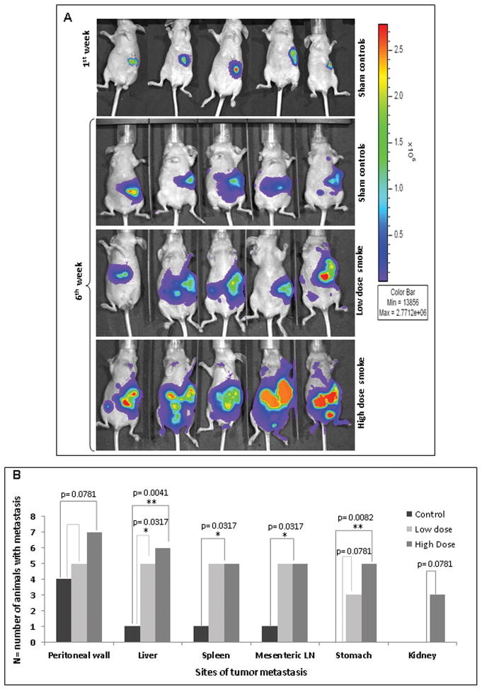
Figure 5.
Cigarette-smoke-mediated increase in pancreatic tumor metastasis 5A. Immunodefficient mice were orthotopically implanted with luciferase-containing CD18/HPAF cells and routinely exposed to an average of 100mg/m3 TSP for the low-dose and 247mg/m3 TSP for the high-dose of cigarette-smoke, for six weeks. The Representative in vivo bioluminescent imaging of the CS-exposed mice showed an increased tumor size as compared to the sham controls and a significant increase in the metastasis in both the low-dose-exposed animals and high-dose-exposed animals as compared to the sham controls. All the images are normalized to the same scale. 5B. Statistical analysis showed significant tumor metastasis to various potential organ sites, including the peritoneal wall, liver, spleen, mesenteric lymph nodes, stomach and kidney. P-values lower than 0.05 were considered statistically significant (*).

