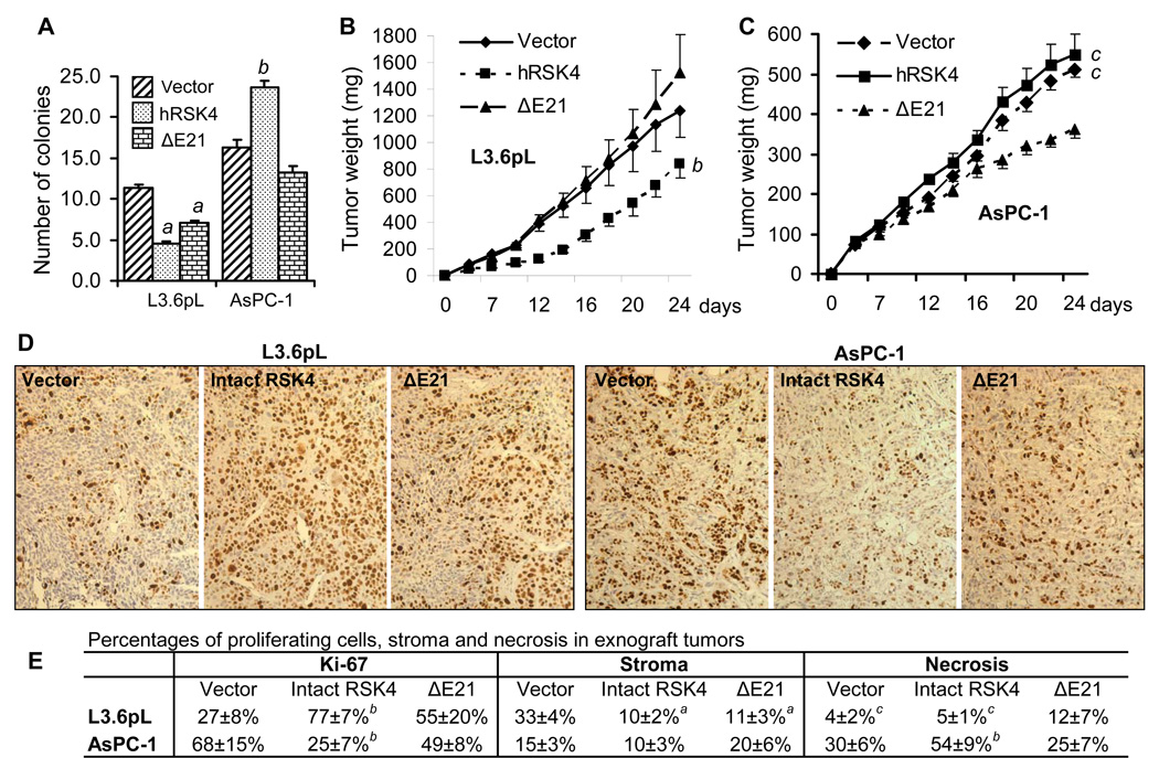
Fig. 11.
Effects of RSK4 on colony formation in soft agar and on tumor growth in SCID mice. Data are presented as mean ± SD. L3.6pL and AsPC-1 cells sorted for the intact hRSK4- or ΔE21-containing pMIG retroviruses were seeded in soft agar or inoculated to SCID mice. A: The colony number of the intact hRSK4 or the ΔE21 expressing L3.6pL cells in soft agar is significantly less than that of the vector expressing cells. On the contrary, the AsPC-1 cells expressing the intact hRSK4 develop a larger number of colonies than the vector or the ΔE21-expressing cells. B: At the last two time points, the intact hRSK4 tumors of L3.6pL cells weigh less than the vector or the ΔE21 tumors. C: At the last two time points, the xenograft tumors of the intact-hRSK4 expressing AsPC-1 cells weigh similarly to the tumors of vector expressing cells but both are much larger than the ΔE21 tumors. D: Representative images of Ki-67 staining of the empty vector, the intact hRSK4, or the ΔE21 expressing L3.6pL and AsPC-1 xenograft tumors. E: Quantification of Ki-67 positive tumor cells as well as stromal and necrotic areas in L3.6pL and AsPC-1 xenograft tumors. a: significantly different from the vector counterpart (p<0.05); b: significantly different from both the vector and the ΔE21 counterparts (p<0.05); c: significantly different from the ΔE21 counterpart, which for the tumor size is at the last two days (p<0.05).

