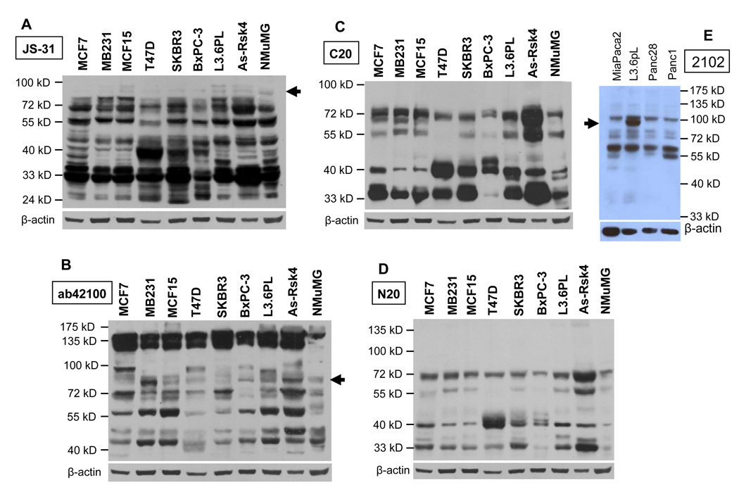
Fig. 6.
Determination of whether endogenous RSK4 proteins are detectable. Proteins from AsPC-1 cells transfected with the pMIG-hRSK4 construct were loaded at a less amount as a positive control. The proteins at 72- and 55-kD are the dominant ones and appear as duplet or triplet in most cell lines, as detected by most antibodies, whereas the putative wt hRSK4 proteins at 90-kD (arrow) is only detected by some antibodies, in some cell lines, or at such a low abundance that can be discerned only when the blots (A and B) were slightly overexposed. Some smaller proteins at about 48, 40 and 33 kD are also detected in some cell lines. The JS-31 antibody again shows high affinity to the proteins of mouse origin in NMuMG cells.

